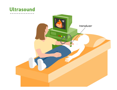
An ultrasound uses sound waves to create an image of the tissue or organ that is being studied. The image can be seen on the screen of the ultrasound machine.
Clear, cold jelly is placed on the part of the body that is being studied. A small round handle (transducer) is then placed on the jelly and moved around. The transducer emits sound waves that bounce off solid parts of the body to create a picture. It can be used to guide a biopsy in real time.

This test itself does not hurt. Sometimes the pressure of the handle on the body can be uncomfortable, particularly if it is placed on an area that is already sore.
